Digital pathology is revolutionizing pathology evaluation across all fields, from human pathology to veterinary diagnostic pathology and toxicologic pathology. The benefits of going digital include faster turnaround times, increased collaboration, improved accuracy with tissue image analysis decision support tools, and reduced costs. With digital pathology, whole slide images can be easily accessed, shared, and analyzed by pathologists and scientists from anywhere in the world, making it an indispensable tool in modern healthcare.
Introduction
Digital pathology is rapidly transforming pathology evaluation across all fields, from human pathology to veterinary diagnostic pathology and toxicologic pathology. The benefits of going digital include faster turnaround times, increased collaboration, improved accuracy with tissue image analysis decision support tools, and reduced costs. With digital pathology, whole slide images can be easily accessed, shared, and analyzed by pathologists and scientists from anywhere in the world, making it an indispensable tool in modern healthcare.
However, for pathology labs, the decision to go digital comes with a major question: what scanner should they use? While the scanner is just a part of the laboratory’s digital transformation journey, it is a crucial component without which the whole process cannot be set in motion. The decision to choose a scanner (or multiple scanners) depends on the digitization approach chosen by the lab.
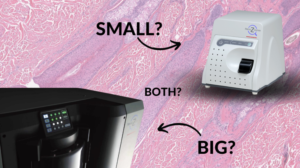
There are fundamentally two approaches to digitizing pathology workflows: “going all in” or “digitizing incrementally.” Going all in often requires one or multiple high-speed, high-throughput scanners, to achieve both the fastest per slide scan time and largest number of slides per scanner at a time. Alternatively, the incremental approach can be initiated with a low-throughput, small-footprint whole slide scanner that may still have fast scan times but has a much lower capacity of slides, making the instrument more compact and simpler.
Labs need to decide on the best approach to digitizing their workflow, and the choice of scanner(s) is an important part of that decision. The adoption of digital pathology is no longer a matter of if, but when and how. Pathology labs cannot afford not to go digital anymore.

Going All-In on Digital Pathology
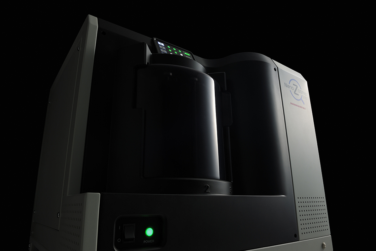
Going all in means that the lab is shifting the entire diagnostic workflow to digital slides, and everything that was previously evaluated on glass will now be diagnosed on digital images. High-volume pathology and histology labs with a high diagnostic or research demand can benefit significantly from this approach and they will require one or more high-speed high-throughput whole slide scanners. These scanners can process hundreds of slides per load and thousands of slides per day and automatically distribute case images to the responsible pathologists streamlining workflow for faster diagnoses.
Digital pathology is expected to improve turnaround times compared to traditional pathology methods. Although the increase in efficiency and decrease in turnaround time depends on several factors such as image management system used, the way the images are accessed (necessity to download locally or not), remote access to images, presence of digital laboratory workflows and automations and the pathologist’s expertise and workload, the scanning speed plays an extremely important role as this is where the digital pathology journey starts. The use of a high-speed high-throughput scanner can significantly reduce turnaround times and save costs.
One key consideration when choosing a high-speed scanner is digital storage capacity. A high-speed scanner generates a massive amount of data, which can quickly exceed the storage capacity of many pathology labs. As such, it is crucial to ensure that the storage infrastructure can accommodate the data generated by the scanner and provide appropriate security and data backup protocols.
The advantage of digital pathology inherently means that images will need to be transferred to different locations for evaluation. Sufficient internet bandwidth is critical in assuring adequate speed and quality of data transmission between image acquisition and image viewing and evaluation.
In human pathology labs, the compliance of the scanner with regulatory requirements must be considered. In the US this includes the necessity for the scanner to be an FDA-cleared medical device.
Incremental Approach to Digital Pathology

In contrast, labs with a lower diagnostic demand can benefit from taking an incremental approach to digital pathology. A low throughput and small footprint whole slide scanner might be sufficient for these labs. These “desktop” scanners are easy to use and can be operated without extensive training. They are ideal for labs that have limited space or do not have a high diagnostic demand. The initial cost of purchasing and implementing these scanners is also lower than that of high-throughput scanners.
Small scanners are also ideal for veterinary labs, which typically have a lower diagnostic demand than human pathology labs. A small scanner is sufficient for these labs and can provide access to pathologists working remotely or in a consulting capacity without the high costs associated with bigger scanners.
Another scenario where a small scanner can be beneficial is if only part of the workflow is using digital slides, such as for expert consults with external pathologists or for education, tumor boards or pathology working groups.
Combining High-Throughput and Low-Throughput Scanners
In some cases, a combination of high-throughput and low-throughput scanners can be an effective option. While a high-throughput scanner is ideal for routine cases, a low-throughput scanner can be used for special stains, IHC or STAT cases requiring immediate pathologist attention, diagnosis and communication to guide the treatment decision. This approach provides flexibility, as there is no need to stop the big digitization machine for targeted or emergency work.
When considering a multi-scanner ecosystem it is crucial to ensure compatibility of the scanner outputs with other tools used for slide evaluation such as slide viewers as well as image management systems. Although not necessary it often helps to have the scanners provided by the same vendor.
Often digital pathology is used both for primary diagnosis and for research projects, which can also require a multi-scanner ecosystem. In the case of primary diagnostic use, it is necessary to use a scanner that is cleared or approved by the applicable regulatory agency for this intended use and conduct an adequate digital pathology system validation.
- In the United States whole slide scanners are classified as Class II medical devices by the U.S. Food and Drug Administration (FDA) and a 510K clearance is required to use them in medical settings. The scanners currently cleared by the FDA include: Philips Ultra-Fast Scanner (cleared in 2017)
- Leica Biosystems Aperio AT2 Scanner (cleared in 2019)
- Hamamatsu NanoZoomerⓇ S360MD Slide scanning system (cleared in 2022)
In addition to the FDA-cleared system, Hamamatsu offers many different scanners for research and pharmaceutical markets which potentially makes the company a one-stop shop for all your digital pathology needs. These scanners provide an efficient and flexible approach to pathology digitization, allowing labs to handle a range of diagnostic and research demands.
In human pathology labs, the compliance of the scanner with regulatory requirements must be considered. In the US this includes the necessity for the scanner to be an FDA cleared medical device.
The Benefits of Digital Pathology
Regardless of the approach taken, the benefits of digital pathology are numerous and undeniable. One key advantage is the possibility to use image analysis algorithms for quality control of the slides and scans as well as decision support tools that have been proven to increase the efficiency and accuracy of pathologists’ work.
Digital pathology also makes it easier for pathologists to collaborate with other clinicians and researchers. Whole slide images can be easily shared and analyzed, allowing for more effective collaboration and better patient outcomes.
Digital pathology also has the potential to reduce costs, especially in settings where the pathology workforce is working remotely and the cost of slide transportation between the lab and the pathologist can be eliminated.
Conclusion
In conclusion, digital pathology is rapidly becoming the new standard in pathology diagnosis, and the question is no longer if labs will digitize their workflow, but when and how. High-volume pathology labs with a high diagnostic demand can benefit significantly from a high-speed, high-throughput whole slide scanner, while labs with a lower diagnostic demand may opt for a low throughput and low-footprint whole slide scanner. Additionally, a combination of high-throughput big scanners and lower-throughput small scanners can provide a more flexible approach to pathology digitization.
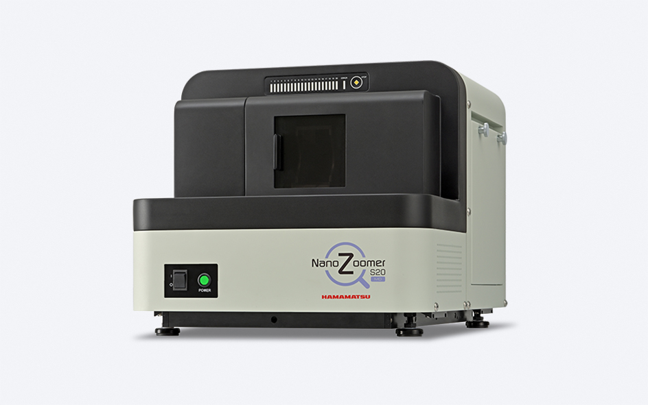
Hamamatsu offers a range of whole slide scanners that cater to a variety of pathology labs, from the NanoZoomerⓇ series with high-speed and high-throughput scanning capabilities to the low footprint NanoZoomerⓇ SQ. Hamamatsu also offers compatible scanners that can work in tandem, such as the NanoZoomerⓇ S20, S60 and the NanoZoomerⓇ S210.


Comments are closed.