


Grundium’s Ocus – the scanning microscope review. This post is a review (with a video demo) of the whole slide scanning microscope Ocus from Grundium. For more information visit Manufacturer: Grundium
What is Ocus and what does it do?
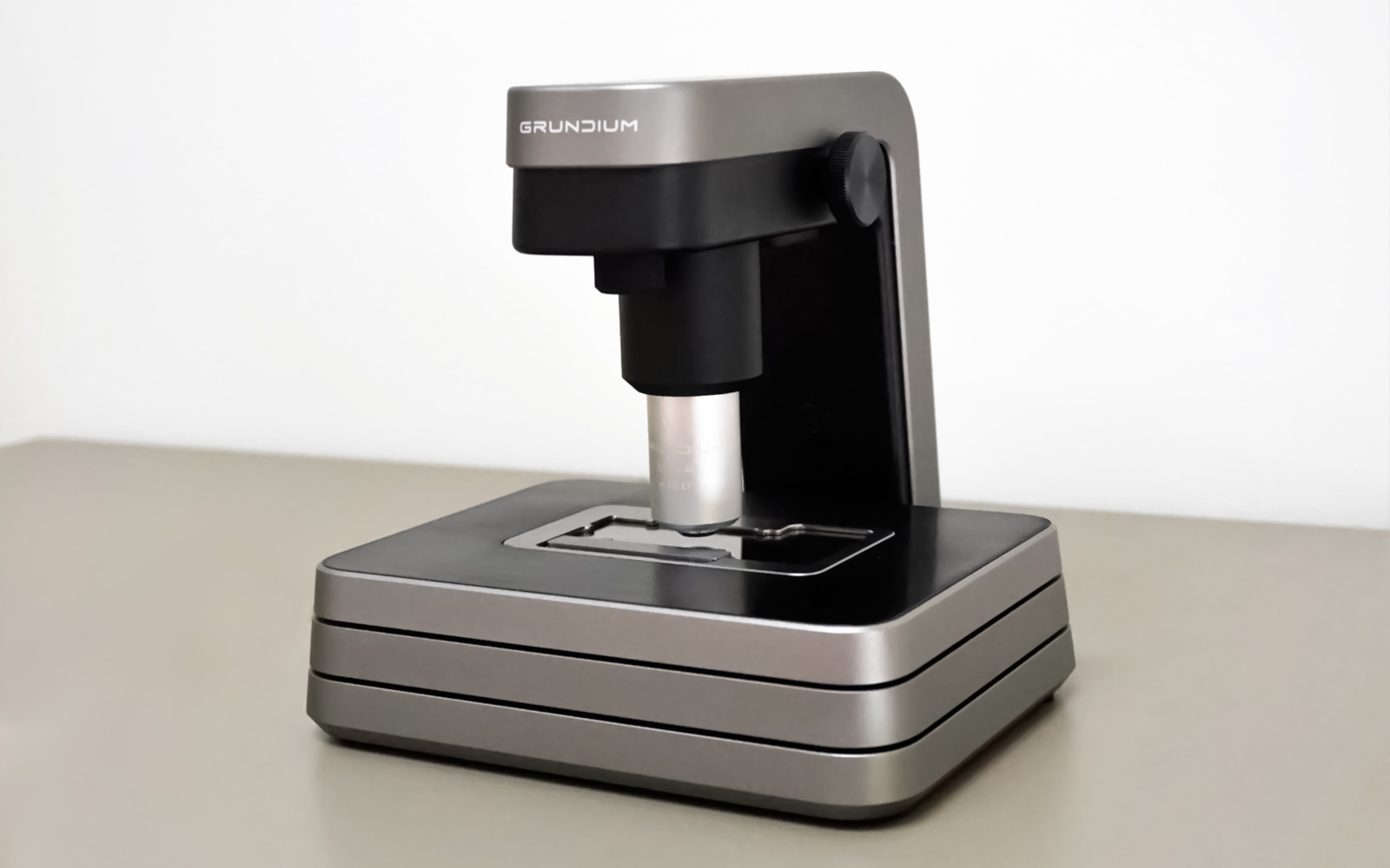
Ocus is a single slide portable whole slide scanning microscope. It enables the scanning and live viewing of one slide at a time. The device connects via wi-fi to a computer or any other device with internet access and a screen and lets you navigate the viewing and scanning process from there. To navigate the device, you use the grundium.net website where you set up an account when you purchase the device. There is no need for additional software installation and the website/ device navigation is very intuitive.
The whole slide images (WSI) are stored on the device and can be exported in SVS, TIFF or pyramid TIFF format and downloaded to a computer or a hard drive. There is an instant slide sharing option either via a link that can be copied and sent to collaborators or through a live online session. In live view mode pictures of single fields of view can be taken in JPEG format.
Technical specifications
The device is light (3.5 kg/ 7.7 lb) and small (18 x 18 x 19 cm/ 7 x 7 x 7.4”) and with the 20x objective scans the slides with a 0.48 um/ pixel resolution and takes ca. 2 minutes to scan a 15 x 15 mm specimen. More detailed technical specifications can be found here
Features and benefits
The small size and lightweight of the device make it extremely portable! It can be put anywhere in the office or easily taken on the road and used in multiple locations.
Internet connectivity makes it an immediate telepathology tool. It can be used for consultations, documentation, and illustration of interesting or relevant cases, education, or any other telepathology application.
How I would use it
My main use of the device would be for remote consultation. As a toxicologic pathologist, I evaluate the effects different compounds and drugs have on animal models. Some compound groups cause well-characterized tissue changes, but others are hard to predict. It is invaluable to be able to ask more experienced colleagues for their opinion and the Ocus would enable me to do it in a matter of minutes from my home office. This is approximately the time it would take me to visit a colleague in the office next-door, only now, I can invite more people from different locations for this digital review at the same time from any location.
Other potential uses
- Remote review of frozen sections
- Central review in multicenter clinical trails
- Pathology consultation from remote locations lacking pathologists
- Digital archiving of interesting cases for pathology rounds
Pros & Cons
Pros
- Portability – the device can be taken and set up anywhere. The only requirement is internet access (a hotspot from a mobile phone is sufficient).
- Modern, minimalistic design – Ocus has just one button, a few ports for different cables, the objective lens, and the slide stage. There is nothing superfluous in this microscope. It is a beautiful piece of personal laboratory equipment.
- Ease of use (once set up) – the communication between the website and the device is seamless and the instructions for the user are easy to follow. There is practically no learning curve – you set it up and start using it from the get-go.
- The quality of the scans is equivalent to any other (larger) whole slide scanner. The resolution is the (more or less) standard 0.5 (to be exact 0.48) um/ pixel like in any other whole slide scanning device with a 20x objective
- The instant sharing feature – you can send links to the scanned slides to your colleagues and have them consult with you on your cases immediately
Cons
- The first setup experience – it was less intuitive than the subsequent use and required assistance from the Grundium team. This might have been caused by the fact that I was not the first user of this particular device, which is not the case when the device is purchased.
- Live view functionality – it could be more dynamic to enable real-time seamless viewing and mimic the experience of a light microscope. It was faster and easier to scan the whole slide, than to review it in the live view mode
Things worth adding
- Cloud storage of the WSI – currently to access the scanned slides, they either must be exported or the Ocus needs to be connected to the internet (the scans are stored within the device). A valuable service would be a cloud-based WSI storing space to back them up and make them accessible when the device is not connected. However different cloud platforms such as Aiforia, Techcyte, and Pathozoom are supported
- Integrated image analysis tools – they are not available yet, but the Grundium team is already exploring different artificial intelligence-based image analysis partnerships and possibilities.
Price range
The Ocus costs a fraction of most multi-slide whole slide scanners. To learn more about the cost reach out to the Grundium team.

Conclusion
This nicely designed, user-friendly, portable whole slide scanner is very complementary to the current digital pathology market. It scans one slide at a time at the speed of…. In the pathology product landscape, it can be placed between the light microscope and a multi-slide whole slide scanner and can be used as a complementary device to both. Scanned slides can be immediately shared by sending a link via e-mail or any other communication platform. It is a great tool for all applications of telepathology including remote diagnosis, remote consultation, interesting case documentation, and archiving and education.
To learn more about getting your own Ocus you can contact the Grundium team here.
Comments are closed.