


If you are planning to digitize your pathology slides and embark on the digital pathology journey, then you need a whole slide imaging (WSI) scanner. However, purchasing a digital slide scanner is a serious financial decision that should not be taken lightly.
This piece of equipment is not something that you will upgrade in two or three years when the next model comes out. This is a purchase that needs to last and keep supporting your lab for the years to come. So, how do you choose one?
In this blog post, we will provide you with a list of 10 questions that will help you define your digital pathology and slide scanning needs. Answering these questions will help you engage with any whole slide scanner vendor on the market to pick the best scanning solution for your pathology lab.
This blog post is based on the Digital Pathology Podcast episode “How to choose a whole slide imaging scanner? w/ Doug Stapleton, Hamamatsu”
Have you been thinking of embarking on the digital pathology journey in your lab? Are you thinking of starting to do tissue image analysis? Then you need a whole slide imaging (WSI) scanner to digitize your pathology slides!
But which WSI scanner should you choose? How do you approach this process? Purchasing a digital slide scanner is a serious financial decision that should not be taken lightly. This piece of equipment is not something that you will upgrade in two or three years when the next model comes out. This is a purchase that needs to last and keep supporting your lab for the years to come. So how do you choose one?
First, YOU NEED TO KNOW WHAT YOU WANT AND WHAT YOU NEED.
Below you will find a list of 10 questions that will help you define your digital pathology and slide scanning needs. Answer the questions and you are ready to engage with any whole slide scanner vendor on the market to pick the best scanning solution for your pathology lab.
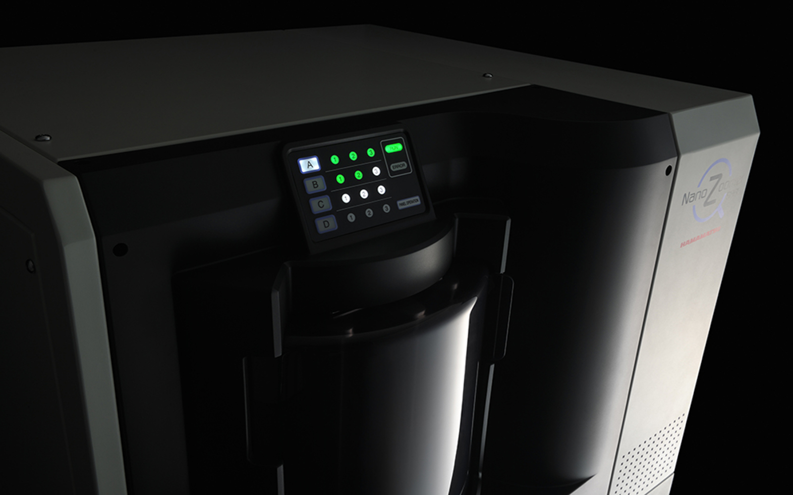
 Whole slide imaging scanner Nanozoomer, Hamamatsu, photo: courtesy of Hamamatsu
10 questions you need to ask yourself before buying a whole slide imaging scanner
1: What is your budget and your throughput?
These are the two main questions you need to ask yourself when you start looking for a whole slide scanner. What can you afford? And how many slides per day/ week/ month will you scan? Those two questions go hand in hand, because your budget may limit the throughput you can afford and the throughput you need can define the budget you must dedicate to a whole slide scanner.
2: What is your intended use now and in the future?
What imaging modality will you be using? Brightfield, immunofluorescence or both? Is it just brightfield images for research or routine diagnostics or are you mainly interested in imaging immunofluorescent stains? Maybe you are working with brightfield images right now, but want to expand your methods in the future? There are options for every scenario – just brightfield, brightfield, and fluorescence or a brightfield scanner with the option to add the fluorescence module in the future.
3: What is the demand in your organization?
Often a WSI scanner is purchased for one department of an organization, but when other members find out about it, the device suddenly becomes more popular than originally intended and the initial throughput is not enough anymore. Be sure to check if there is demand for whole slide imaging outside of your department and think about how the demand may change over time.
4: How much space do you have in the lab for your scanner? Where can you put it?
To ensure uninterrupted scanning and the highest quality whole slide images, the whole slide scanner needs to rest on a stable countertop protected from vibration. It is important because the vibration caused by lab equipment, such as centrifuges and freezers, makes a scanner’s optics shake while scanning. Make sure that the scanner is far enough away from these kinds of devices.
Whole slide scanners usually need additional hardware such as a dedicated computer. Think about how you can carve out a small working area for all the necessary equipment around your scanner
5: What size of pathology slides do you want to scan?
Are you working with the traditional 3″×1″ (75 mm × 25 mm) slides or are your specimens bigger? Find out in advance if the WSI scanner you are thinking about can handle different slide sizes.
6: What magnification will you be scanning at?
What is the maximum magnification you want to be able to scan your slides at? Is 20x enough, do you need 40x or maybe more? Are you working with cytology specimens? If so, check if the WSI scanner is capable of Z-stacking (scanning the samples at multiple horizontal planes).
7: Is it just the WSI scanner or does it come with additional equipment?
Because you will need more than just the WSI scanner itself to interact with the digitized pathology slides you need to check if the scanner you are thinking of comes with additional equipment such as:
- slide viewer
- high-resolution monitor
- mouse
- image management system?
If it doesn’t you will have to take care of those parts of your digital pathology system on your own. Whether it is a good or a bad thing depends on your individual preferences. Be sure to compare e.g., different slide viewers and image management systems in advance.
8: How do you want to interact with the WSI scanner?
Do you want your scanner to be automatic, hands-off, where you load it with slides and walk away so it can do the scanning while you work on something else?
Or would you rather have it semi-automatic, where you load it, view the slides, and pick your scanning regions?
Or another way would be a single slide mode, to just scan one slide at a time. This is the most cumbersome option but also the one that gives the most flexibility regarding the times of scanning.
Which one do you need or prefer?
9: How easy is it to scan the slides?
Is the user interface of the scanning software intuitive? How many clicks does it take to initiate the scanning process? This may not seem so crucial, but it is important because life in the lab can get really busy so anything that streamlines and accelerates the scanning process helps
Hamamatsu offers all of them which potentially makes them a one-stop-shop for all your digital pathology needs.
10: How does the whole slide imaging scanner integrate with other systems in your lab?
Most likely before bringing a digital pathology system onboard, many other systems, including a lab information management system (LIMS) will already be in use in your lab. Before purchasing a WSI scanner, make sure there is a good way to integrate it with other systems in your lab. This can happen through an open application programming interface (API) with the vendor’s assistance or with the assistance of your own IT department.
Bonus Question 11: Are there any extras included in the package?
Is there a special functionality that you are interested in included in the deal, such as:
- telepathology capabilities
- virtual slide conferences
- slide annotation capabilities
- or cloud storage options?
Hamamatsu offers all of them which potentially makes them a one-stop shop for all your digital pathology needs.
The questions above will help you define your needs regarding a digital pathology system. Equipped with the answers you can talk to your colleagues who already embarked on the digital pathology journey or contact any vendor and start your “WSI scanner shopping”.
Just remember, no matter which WSI scanner you choose, always dry, and clean your slides before scanning!
Comments are closed.