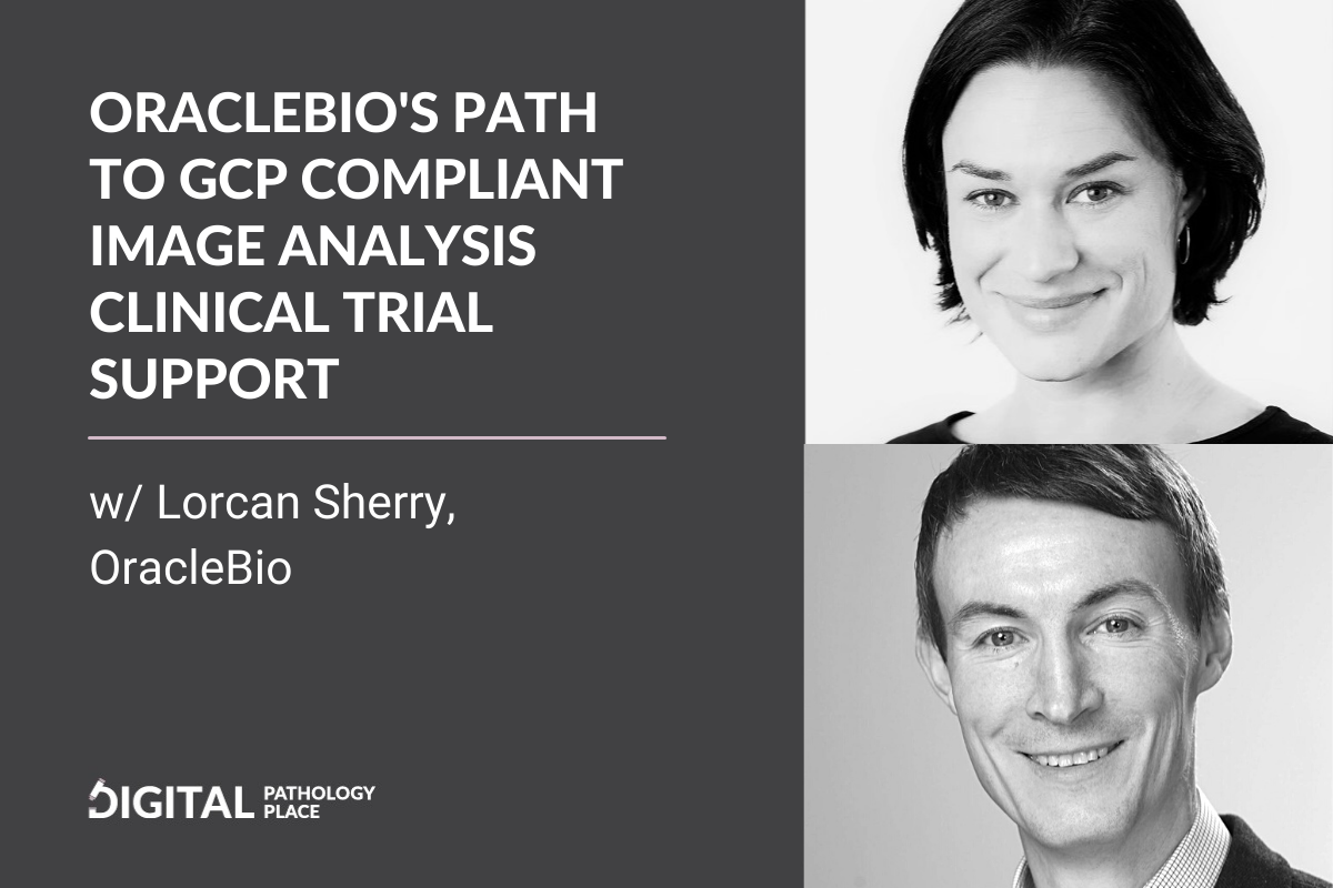
Aleksandra: [00:01:08] Today my guest is Lorcan Sherry from OracleBio. Hi, Lorcan. How are you?
Lorcan: [00:01:12] Hi, Aleks. I’m fine, thanks.
Aleksandra: [00:01:14] So let’s start with your introduction. Tell us about yourself, about your background, and how you ended up at OracleBio?
Lorcan: [00:01:21] Sure, sure. And thanks for the invitation to speak with you today, I’m delighted to be here. My background is I’m a pharmacologist by training. I did a degree in pharmacology at the University of Edinburgh, and when I finished that degree, I was then able to start a PhD at the same university in cardiovascular medicine. That gave me some exposure and training in the areas of histology and pathology, with respect to tissue processing, tissue sectioning, immunohistochemistry staining and the use of microscopes to review the staining, and some image analysis techniques. So that was really my real starting point for getting involved in pathology and histology, and I really enjoyed that experience through my PhD.
[00:02:06] When I came to the end of my PhD, I [00:02:08] felt that I wasn’t really wanting to continue in academia. I applied for a job in pharma, a local pharma R&D company, and I was lucky to get a job there working in the histology group to support preclinical cardiovascular projects. And I spent the next 10 years of my career in pharma R&D, where I managed that histology group and supported a range of different types of studies along the R&D pipeline, such as preclinical translational projects, and I got more involved in biomarker research and also biomarker strategies to support R&D projects as they move from preclinical through to clinical. That was a really a great time for me, and I enjoyed that work.
[00:02:48] About 10 years into that, the company itself was acquired by a larger pharmaceutical company, just at the end of 2010, and the company site was closed. So myself and a colleague who I’d worked with extensively through my pharma career, John Waller, John convinced me that we could diversify our [00:03:08] careers and start a CRO in biomarker services, which was an interesting concept for me at the time. I have to say, I think John was more the entrepreneur than I was in that situation, but I thought, “Yeah, this sounds good. Let’s try this.” And from that, OracleBio started, just at the beginning of 2011.
Aleksandra: [00:03:24] Okay, wow. Huh. And CRO for image analysis, this is a pretty unique value proposition.
Lorcan: [00:03:34] Yes.
Aleksandra: [00:03:35] I don’t think there is any… Some companies provide services, but a CRO that is strictly for image analysis is something different. And tell me about this value proposition, like how did you come up with that idea?
Lorcan: [00:03:48] Yes. I’d like to see we were visionaries in the field-
Aleksandra: [00:03:51] you were.
Lorcan: [00:03:51] But at the time, around 2011 when we started the company-
Aleksandra: [00:03:55] oh, in 2011 you definitely were.
Lorcan: [00:03:57] It was more a necessity. We started with the mindset that we would work in the area of biomarker research. We really felt, at the time, translational biomarkers were having a big impact in the R&D [00:04:08] pipeline from preclinical through to clinical studies, and building the confidence level of data that went from preclinical studies through to clinic with more success. So we certainly wanted to do something in that biomarker field. When we sat down and really thought about it, that’s a big area, so we really needed to focus on one particular part of that process. And I think our backgrounds of histology image analysis working in pharma R&D led us to a decision that pathology and image analysis was an area we wanted to focus on. And that’s really the reason that we started, it was really based on our background and our skill sets that we had at the time.
[00:04:45] And it was quite interesting, when I think back about what image analysis was back then, whole-slide scanning was just starting to come into play. I wouldn’t say that image analysis was one dimensional at the time, but it was certainly handcuffed to working with fields of view on microscopes, and maybe working on single chromogenic staining. And it certainly had a purpose and a lot of people were doing this, histology was around for a [00:05:08] while, but image analysis really didn’t have a chance to express itself, and I think that really has happened over the last sort of five to 10 years.
[00:05:16] In the early years for us, it was the advancement of moving from microscope field-of-view images to whole-slide images was a big step forward. Now we were able to generate data from whole sections, as opposed to subjective field of views or smaller regions, and that was sort of a transformational stage. At the same time, histology was progressing very rapidly. It was moving from chromogenic staining to multiplex staining to in situ hybridization techniques. Fluorescent staining, going from two-plex to four-plex to six-plex to eight-plex. All this was advancing, and this was again allowing image analysis to express itself better with these more complex staining techniques and images that we were receiving.
[00:06:00] And then of course, the company, as a company we’ve worked across all of the R&D pipeline and across all [00:06:08] therapeutic areas, but oncology and cancer tissues were a principle part of the samples we were receiving, a majority of the work that we were receiving. And when immunotherapy and immuno-oncology came on the scene around 2013, 2014, this was a real drive in the R&D field. And immuno-oncology really led itself to allowing digital pathology to add real value to that area, because immuno-oncology involved different cell populations in tumor tissue, it involved the immune cells, and clients were now wanting to know about the phenotyping of those immune cells. Are they in the tumor? Are they in the stroma? What other cells are around them, such as macrophages, dendritic cells, NK cells? What’s the spatial relationship of these cells in the tissue?
[00:07:00] And this really allowed image analysis and digital pathology to help answer those questions, and I feel we really got a huge lift from a lot of [00:07:08] things coming together around that time. And that progress has actually continued right up to today with the advent of things like artificial intelligence, image mass cytometry techniques, where clients are seeing that there’s an advancement in the techniques and the quality of the images, and they’re now able to get much more data from their samples than we would have got five or 10 years ago when we started. So I think when we started, we were in this area of, we are helping, but it was single chromogenic answering quite straightforward questions, but now we’re able to answer much more complex questions. And that’s been really interesting, and a really rewarding sort of part of being on this journey with the company.
Aleksandra: [00:07:55] So it’s 10 years since 2011. And definitely now, like you say, immuno-oncology kind of requires image analysis. You cannot do it manually or visually. You can [00:08:08] try, but it’s never going to have the level of consistency, the level of granularity, so-
Lorcan: [00:08:14] absolutely, absolutely. And because we’ve seen some of the histology techniques advancing, they are now being applied to other therapeutic areas. We do a lot of work in inflammation and fibrosis, and we do a lot of work in dermatology. And we are now thinking about maximizing the data that we can extract from images, and we learned a lot from immuno-oncology and immunotherapy and the approaches and the cell types and how to stain them and segment these cells. So I think we’re now being able to apply that learning to other therapeutic areas, to the advantage of generating more data to support better interpretations of those samples.
Aleksandra: [00:08:55] So you’re a CRO, so you are using tools of different companies, and this is something that interests me a lot. You’re using image analysis software from wherever you [00:09:08] think is going to suit best your needs. How do you choose the software? And the second part of this question is, why did you go this route? There are so many companies that decide to do software, and then they also provide services. You decided you’re not going to do software, you’re going to just do image analysis and use what’s out there. Tell me about your vetting process of the software?
Lorcan: [00:09:33] Yeah, yeah. I can tell you a little bit about how OracleBio work, because maybe it helps understand why we’ve taken the approach. So principally, OracleBio provides digital pathology services to support pharma and biotech R&D. So essentially, we receive images from our client’s histology studies, or we receive their slides and we scan those two images, and then we use commercially available software to generate [00:10:08] algorithms tailored to the studies to quantify the staining on those samples. And we generate data from the algorithms, from the images, and return that data to our clients to allow them to better interpret the outcome of their studies.
[00:10:23] We, because of our background, we really tried to think about how we could use the skillsets and experiences that we had in the best possible way to support our clients. We didn’t come from a programming background or an IT background, so we didn’t have that type of internal capabilities when we started the company. And at the time there was some great softwares on the market, and actually our first software that we worked with was Definiens. I think we spoke to Definiens in early 2011 and managed to convince them that it would be a good business model to have CROs using their software, as well as them selling it directly to clients. And by having a software that was powerful like that, [00:11:08] and was on a maintenance contract, we didn’t have to worry about updating it, it allowed us to focus on our skills of… Which is really, partly, a consultancy service that we do for our clients.
[00:11:18] Yes, we receive their images. We use our expertise in image analysis to generate the data, but our clients are quite different. We work with small biotechs right through to large pharma, and some of our small biotech clients will not necessarily have internal histology expertise. They may only do four or five, six studies a year, but they need quality data from those samples. So in that instance, we would speak to our clients and we would say, “What is the question you’re trying to answer from your samples?” And then we can suggest or recommend a histology approach or staining required to best deliver that data.
[00:11:56] So we work with our clients at that level, even before they’ve generated the images in some instances. And our clients may say to us, “Okay, that’s great, can you help with the histology?” So we actually have a network of about [00:12:08] six histology companies that we partner with in Europe, and for quite a number of studies, we manage the process for some of our smaller biotechs from tissue to data. We partner with histology CROs, tissue companies, and we also recently have worked with bio-informatics companies, brought bio-informatics companies into our partnership to be able to deal with the data at the backend and run bioinformatics, all under a contract that we manage.
[00:12:31] So that in itself was a business model that we felt didn’t necessarily need to have our own IP with respect to the software. The other component of that is what we found, although we started with the Definiens software and we were very much on that software as a company, a single platform, and it was a great software, we found that, especially our larger pharma clients, those companies don’t just use one software for all of their image analysis needs. They will use a different range of softwares, and there’s a lot of great softwares out there.
[00:13:00] And there’s a lot of different software, a lot of great softwares out there. So we were going into companies and we were finding that they had different softwares that they were using. [00:13:08] And a client may actually want us to use a software that they’re using internally. So it became important for us to be able to broaden the types of softwares that we were using to align with what they were using internally, and to be able to return the data in a way that they could manage. Or they could send us some algorithms from their internal group and we would run them and then pass the data back. And so that was a flexibility that being software agnostic allowed us to have. And I think that’s probably served us well as we’ve moved forward, because you talked about how do we choose the software for a particular study?
Aleksandra: [00:13:42] Yes.
Lorcan: [00:13:42] Actually, for the first four years we were with Definiens and then that finished, Definiens business model changed, as we now know, and we then acquired software from Indica Labs HALO platform. And then after that, we also acquired Visiopharm’s software. So at the moment we principally run two software packages and they are probably the two most well-known and most used softwares in pharma, R and [00:14:08] D, which is Indica Labs HALO and Visiopharm. But the model does allow us to look at other softwares moving forward and there’s other softwares like QuPath out there.
Aleksandra: [00:14:17] That what I wanted to ask you about, if you incorporate this in any way.
Lorcan: [00:14:22] Yeah. And that’s something that we are looking to do as we move forward, because that’s a software that is becoming much more used by a lot of our clients and things like MATLAB and programming capabilities as well. I think when we have that wider menu board of softwares to work with, that it makes us more flexible in how we can work with clients, but also thinking about which software solution best suits the study that we’re working on. And every software has its strengths and weaknesses. I don’t particularly want to go into them right now, but-
Aleksandra: [00:14:54] everybody who starts using is on their own.
Lorcan: [00:14:57] But certainly for studies, we will first have a conversation with the client, understand the needs of their study, understand the data readouts that they want to generate that are important. And then [00:15:08] we think about which software is best suited from the softwares that we’re using to allow us to generate that data. Again, that flexibility is important for us as a company.
Aleksandra: [00:15:18] Tell me about a failure that sets you up for success. Did you have anything that , you thought it was a disaster, but then later, you came out stronger on the other end.
Lorcan: [00:15:29] Yes. Yes. That’s a good question. Yeah I don’t think we’ve had any sort of catastrophic failures in the company that we’ve had to pick ourselves up right from the ground and start again.
[00:15:39]But I guess in the early years we were using software from Definiens that we leased from Definiens on an annual basis. And and it was a very powerful software and it really allowed us to do image analysis well on the samples we were receiving. But after about four years we weren’t able to continue.
[00:15:57]That lease period, Definiens changed, their business model and politely told us that, they would not want to continue. And at the time we, this was our principal software and we were like, okay, this is not [00:16:08] good. You know, how do we get out of this? And, luckily at the time Indica Labs had started off maybe a year or two before, and they were making some really nice progress with HALO and we spoke to them and we were able to acquire a large number of licenses of HALO that we owned.
[00:16:23]And, and that actually dovetails very nicely with the sort of the winding down of the Definiens lease period. And, and from that we learned the lesson that maybe that we shouldn’t just be a a single software company. I think flexibility with respect to just business continuity is good.
[00:16:38] And, And from that, we brought on, we, we purchased Visiopharm software a year or so later and built up that platform in the company. And we are, uh, open and active in exploring other opportunities to bring in other softwares that, that allows us to, to widen that sort of menu of all of platforms.
[00:16:57] And I think that was. At the time we probably saw it as a failure that we couldn’t convince Definiens to continue with us, but looking back, there was other things. It wasn’t, I don’t think it was just about us. I think there was a business [00:17:08] model that Definiens were pursuing that didn’t necessarily you know, Yeah, allow them to do it, to at least software to CROs.
[00:17:16] But but it certainly, it was one step backwards, but two steps forwards. I think, if we look where we are now and Definiens is no longer on the market and we have two very powerful softwares we have flexibility to bring on more that’s big thing I think has been a real benefit for us.
Aleksandra: [00:17:31] Now, the market is also, a little bit more full of digital image analysis software.
[00:17:37]So you mentioned you want to provide quality data. My question is, how do you provide the quality? What’s your process for assuring that the quality of your data is high? And I’m meaning just the quality of the image analysis markups, that it matches what’s in the tissue image?
Lorcan: [00:17:59] Yeah. Yeah, this is a big question and there’s lots going on at OracleBio about how we address and [00:18:08] embrace quality in our processes. Maybe just if I can take a little step back when, as the company has evolved and we’ve grown over the last 10 years, we certainly do work across the R & D pipeline. As I said, in pre-clinic target discovery, preclinical translation, and clinical, but actually over the last two, three, four years, we’ve become more focused on clinical work. And that’s where, when I say that, I’m talking about clinical trials where histology has been used in clinical trials and mainly oncology clinical trials and clients are looking for exploratory biomarker data to support some of their end points. But we also have found that clients are also looking for data to be generated to a quality standard that could then be used as part of a data package for submission, for regulatory review. Okay? A couple of years ago at OracleBio, we started the process of incorporating GCP or good clinical practice into our workflow. Our workflow at the [00:19:08] time was still quite robust.
[00:19:10] So for example, from receiving an image, we will receive in a set of images for a study. We first perform a QC of every image that we receive, and that’s a combination of visual accuracy with respect to tissue. Is there enough tissue present in the image? What are the artifacts? How is the staining tissue folds? Things like that, do they need to be removed? We do a general quality assessment of the tissue. And we also look at the quality assessment of the staining, which can be used with some algorithms looking at the foreground and the background levels of staining to check if any of those sections are overstained or understained and so things. So from that process, only images that pass our QC process then go into the algorithm stage and the algorithm development stage.
[00:19:54] When it comes to algorithm development, there’s really two parts to this essentially. In the most basic terms, there is a segment segmenting your tissue into regions of interest that you want to perform your analysis in. So for [00:20:08] example, a tumor sample may have some normal tissue or healthy tissue present, and it’ll have tumor present. And we use pathologists in our company to outline where the tumor microenvironment is present on those sections to exclude non-tumor tissue. And then within that tumor microenvironment, we develop first a classifier and an algorithm to segment that tumor microenvironment into tumor tissue and stroma tissue. And we may remove things like necrosis and then regions of glass or white space. And then when we’ve segmented the tissue, we create a second app, which then looks at the cells present in those regions of interest or the staining, and we quantify the staining with a second app. Now through that process, I mentioned we use pathologists to start with. So not only do the pathologists annotate our tumor microenvironment, but they also help validate the algorithms that we’ve generated with our image [00:21:08] analysis software.
[00:21:08] So for something like tumor stroma segmentation, we can then create region, create fields of view on samples. And we can independently have our pathologist annotate where the tumor and stroma is as a gold standard. Ground truth, sorry. And then we will run the algorithm separately on the same field of view. And we will do a dice coefficient scoring to compare the ground truth by the pathologist with what the algorithm has generated. And it has to reach a certain level of criteria for that to be accepted as an algorithm for use in that study. And similarly so for cell analysis, we generate an algorithm to pick out, for example, positive and negative cells for a particular biomarker. Then our pathologist will review the sections in field of views, count the numbers of positive or negative cells or whatever the staining may be. And then we do a correlation of the image analysis readout with the pathologist readout.
[00:22:02] So we have validation steps through that work process, [00:22:08] through to the validation of the algorithms before they’re applied to the images to generate the data. So that has always been there, but GCP is something quite different. And that actually, it was a bigger commitment, but for the company to be able to generate data to GCP requires certain quality standards in the company. And it requires the data to be traceable and evidence of all the steps used to generate the data. It requires the data analysis to be run on validated systems. It requires your organization to be set up in a way that has all its procedures and processes outlined in clear SOPs risk assessment documentation. It’s been a huge undertaking for a company of our size to do this. And it has literally taken us nearly two years.
Aleksandra: [00:23:00] How big are you? What’s your size?
Lorcan: [00:23:01] We’re 20 people now.
Aleksandra: [00:23:02] Oh, congratulations.
Lorcan: [00:23:04] Yeah, so it’s going well in that respect. [00:23:08] But we perform our first GCP study in February this year. And we’ve been audited a number of times. And that’s an inflection point for the company. We’re very excited about this. It’s something we’ve been working towards. And we truly believe that being able to perform image analysis to GCP and bringing that data to a quality standard and allowing the data to be reviewed, to support a regulatory submission moves digital pathology forward in that respect. And we’re working in the pharma R&D environment. We’re not necessarily in the diagnostics field, so we’re not generating diagnostics. This is data that will support a therapy moving through to the market. And I think, as you said, we’re not aware of other companies doing this.
Aleksandra: [00:23:54] I’m not aware about other companies doing this in that way either. First of all, congratulations on this. This is a huge step and you anticipated my next question. How do you approach [00:24:08] the regulatory requirements in the pharma industry?
[00:24:12] And this is big, being GCP compliant. And I also ask this question because we have QuPath, anybody can buy image analysis software, you buy your software, so anyone can do it. How do you, and you explained that, how do you provide the quality? What do you do to provide it, to make sure that this is quality data? And with being GCP compliant, that’s-
Lorcan: [00:24:36] it’s actually… Yeah, it’s a very interesting question, and it’s something that we’ve had to look… Our Clinical Operations Manager, Alison Bigley, had previously worked at AstraZeneca in the UK for 25 years within the digital pathology field and pathology. And Alison was involved in the implementation of the GCP process within AstraZeneca, so we were not coming into this process blind to what was involved.
[00:25:03] It’s different implementing and building GCP in a company like AstraZeneca, than [00:25:08] to a company like OracleBio, but the software validation, and how you validate the software to ensure the data integrity, is actually, it’s a crucial part of the process, but it’s still a small part of the process.
[00:25:19] GCP for us is actually something that impacts the overall company and how we work as a company. The standards that we have to achieve, the… as I said, we have over 200 documents now with respect to standard operating procedures that cover everything from how we approach studies, how we deal with data, how we manage data. But even the IT infrastructure, the servers that are holding our software that are validated, but the disaster recovery processes that need to be put in place, if everything got lost, that all has to be covered from a GCP level. Business continuity needs to be… and actually to study management process, and how we can be audited, and how we collect information and so forth.
[00:26:00] It’s been a real learning curve for the company over the last two years, and we haven’t approached this lightly, and that’s the reason it’s [00:26:08] taken us this amount of time to get everything in place that we feel was required to do this correctly. And of course, once you’re at that stage, then clients will audit you to you to review your processes. And only then, if they accept that your processes are in place, will they work with you as a company.
[00:26:27] It’s been great that we’ve been able to go through our first audit and come out of that successfully, and then move to the study phase. But it’s certainly a bigger process than just the software. So you’re right. Anyone can get access to the software and validate the software, but it’s the actual, the overarching aspect of GCP across the company and how we operate, as well as the software that’s actually as much a body of that effort.
Aleksandra: [00:26:54] You said pathologists are helping with image analysis annotations, with both a visual QC and a QC with automated tests and annotations. How else do they work with you? Or do [00:27:08] you have any other ways you work with pathologists?
Lorcan: [00:27:13] Just to give you a little bit of background of how we’ve interacted with pathologists as the company has evolved…
Aleksandra: [00:27:17] Do you have any pathologists on staff?
Lorcan: [00:27:19] Yes, we do. We have two clinical pathologists on our books. And about up until two years ago, we worked with a network of pathologists, so remote pathologists that were not part of our company, but were in a partnership with. So, if we had a study that required a pathologist review or annotations to be performed on samples, we would tap into our network, which was either some professional companies or our local hospital had a number of pathologists.
[00:27:48] However, I think, a couple of years ago, it became really clear to us that we needed to have pathologists internally in the company. One aspect of that is a credibility aspect. We’re a digital pathology company. We are image analysis scientists. We’ve looked at thousands of images, but we’re not trained pathologists. So there’s a lot of things that we can [00:28:08] recognize on tissue sections, but there will still be a certain amount of things we go, “We’re not exactly sure if that’s a tumor cell or not.” And having trained pathologists work side by side with us has been fantastic in that respect.
[00:28:19] And not only are they annotating samples and helping us review our classifiers, but we’re actually having educational sessions with our pathologists to train our staff. So, for example, yesterday, we had an hour review of gastric cancer, and how the cancer presents itself in different ways, and where it presents itself and how it differs between well-differentiated and poorly differentiated cancer. And actually, these are critical components of how our image analysis scientists then use that information to interpret sections, and support the annotation of sections, the training of algorithms to accurately segment these regions in tissues.
[00:29:00] And I can’t see a time that we wouldn’t work with pathologists internally moving forward. I think it’s a critical component, and it’s [00:29:08] something that we’ve all really enjoyed in the company.
Aleksandra: [00:29:10] Mm-hmm [affirmative]. I think it’s something that all image analysis companies are understanding now. I think, even a couple of years ago, let’s say four years ago, it was not a given. Now, like you say, the aspect of credibility is very important, and this is a certain training that is needed for digital pathology, so I’m really glad to hear that you have people on staff, and they’re working with you closely and they’re bridging the gap in expertise.
[00:29:38] So, another question. How do you innovate? I know you have… So you have the software packages that are there, and you get the updates of the software, but how do you look for expanding? Expanding in the sense of where to go next in digital pathology?
Lorcan: [00:30:01] I look at innovation as being, there’s a practical innovation, which is keeping up with the technologies, [00:30:08] keeping up with the infrastructure, and then there’s a strategic innovation around what is the direction of digital pathology, and what direction do does OracleBio go in that area?
[00:30:17] Maybe from a practical sense first, you’re absolutely right, the softwares are advancing at a rapid rate, especially the Visiopharm and HALO packages that we’ve worked. I think both those companies have done a tremendous job in keeping pace with the requirements of digital pathology, and how both of those softwares have innovated and expanded and grown over the last five or 10 years is remarkable, really, and the implementation of deep learning and AI as well.
[00:30:49] As CRO, we need to keep on top of the software because our clients are obviously wanting to be able to get as much from their tissue sections, and as the software advance, we need to be experts in the software, and that is a job in itself.
[00:31:02] We also need to be on top of the advances and innovations in histology, and I talked about, [00:31:08] earlier, about how histology has gone to multiplexing, from single plexing to multiplexing. And now we’re working with cancer samples that have stained with four, eight or 12 different markers. And then how do we analyze those types of samples, and how do we deal with the data that comes out of those types of samples? And that’s an innovation in itself. Thinking about how we approach that is important for us to deal with.
[00:31:32] The third practical example I would give you is working with the infrastructure required to power and run these softwares. And we, about 18 months ago, diversified from working directly in a data center, but also now we have software in AWS, which gives us immediate scalable access to CPUs and GPUs.
Aleksandra: [00:31:58] AWS, Amazon WorkSpaces, just to clarify.
Lorcan: [00:32:00] Sorry, yes. Amazon, yeah. And it gives us a realtime scalability for our services. And especially if [00:32:08] we’re working with deep learning algorithms where we need GPU access, we can have a hundred GPUs or one GPU with a flick of a switch. So that in itself has allowed us to really work on bigger studies and build that sort of environment to future-proof our scalability and that respect moving forward.
[00:32:26] Strategically, strategically, there’s so much happening in digital pathology, Aleks, that it’s… The runaway scientist in us feels like we want to do a bit of everything, but the businessperson in us thinks that we’ve got to focus somewhere, because you spread yourself too thinly, then it becomes very difficult to keep a pace, and to be successful in all these areas.
[00:32:47] I think, for us, we’ve looked strategically at that area that I talked earlier about, clinical trials, data from clinical trials. How can we move that data from being exploratory biomarker to something that could actually be used in regulatory decision making? And we believe GCP is a step in that direction.
[00:33:04] There’s also the thought of the algorithms that we are developing [00:33:08] for some of these studies could potentially be used for companion diagnostics in the future. So there’s opportunities to really think about how we could help our clients who are performing clinical trials, and finding particular readouts or groups of readouts that are able to stratify patients for responders, non-responders. Could that be used as a way as a companion diagnostic in the future? And that’s a really interesting and exciting area as well.
[00:33:38] And I see that slightly different to maybe the healthcare diagnostics for deep learning has been used at the minute, but there’s a lot of great companies working in the healthcare environment, where they are creating a deep learning app that will be used by a pathologist to support decision-making for a diagnostic purpose.
[00:33:55] Whereas we are not necessarily in the healthcare diagnostics environment, but we’re more in the later stage R&D clinical environment, supporting the generation of quality data, but also potentially [00:34:08] working with our clients to identify readouts that could be built into an algorithm that could help identify patients who would respond to that therapy, and be used as a diagnostic prognosis to identify the patients who will respond to that therapy.
[00:34:23] So the short-term goal for OracleBio is really to focus on GCP and build that, but also to continue to develop the relationships we have with small biotechs. They are the guys that we start with in preclinical, and they stay with us as we work through the R&D pipeline, so they’re very important, that we don’t lose sight of, working with the smaller companies or the smaller projects in the preclinical environment. But really, I think we’ve got an opportunity in the later stage clinical environment to hopefully make a difference.
Aleksandra: [00:34:55] Both of the software packages you mentioned have AI and deep learning modules, and you said you’re using it. How did it change your image analysis process? What can you do now that you couldn’t do [00:35:08] before? And also, another thing, do you combine approaches? And how much of your approach is still the classical computer vision rule-based image analysis, and how much is has moved to the AI?
Lorcan: [00:35:21] Yes, good question. Because again, in the area that we’re working on, we see deep learning slightly differently, the deep learning algorithms differently to, say, if you were working in diagnostic healthcare. So, in diagnostic healthcare, you would be creating a product, which is your deep learning algorithm.
[00:35:40] Our product, at the research level, at the R&D level, is data. And actually, the deep learning algorithms are being used to support the generation of data. We have deep learning modules for both our HALO, Indica Labs HALO platform, but also, our Visiopharm platform.
Aleksandra: [00:35:57] I think QuPath has incorporated for sure, machine learning and I think some deep learning options, as well.
Lorcan: [00:36:04] Absolutely. It has [00:36:08] been transformational. Previously, there are types of studies where a client would have asked us to quantify a biomarker in tumor and stroma of oncology sample. But to segment the tumor and stroma, we would have needed a serial section that would stain for a tumor mask, like an epithelial marker or something like that, cytokeratin. We would have created the outline of the tumor and stroma on the serial section with the cytokeratin highlighting the tumor. Then, we would have co-registered with the biomarker section and transferred the annotations to the biomarker section to analyze within those regions. It was a bit more of a protracted approach but the reason we had to do that was because the classical machine learning techniques were just not powerful enough to segment a tumor and stroma on the biomarker section alone.
[00:36:57] What deep learning has allowed us to do is, to do exactly that. We’ve been able to train the algorithms directly on biomarker stain sections, and it was [00:37:08] segment accurately, the regions of interest, so that we don’t need a separate section to support that anymore. That’s been quite critical, especially in clinical studies where clients don’t necessarily want to stay in a number of extra sections of their sample to just be able to find where certain structures are. In that instance, it’s been very powerful.
[00:37:31] I think what we’ve learned over the last two years of using deep learning in our environment is that it’s still a challenge to understand when best to use deep learning and when to use classical machine learning because the studies that we’re working on, sometimes they can be a hundred samples and the question is quite specific to those samples. If we needed for machine learning apps to pick out the regions of interest across those 100 samples, or we need one deep learning app, but the one deep learning app may take numbers of days to annotate, to train, further train, and then you have it working, but it’s taken about a [00:38:08] couple of weeks to do that. You get data from your study eventually, but that deep learning app can no longer really be used for another study because the next study that comes along is a slightly different tissue sample or tissue type and different staining.
[00:38:20] It works very nicely in say, safety tox studies, where the types of staining approaches are quite consistent. All that effort of building your deep learning app at the start, you get the benefit with repeat studies. But in our environment, we don’t necessarily see so many repeat studies because they’re coming in from different clients and different tissue types and different staining techniques and they’re answering a particular, or a specific question to those samples.
[00:38:45] We do need to be slightly careful about how much time and effort we put into developing deep learning versus classical machine learning apps but I think as we move forward, we will embrace deep learning more and more. And actually, I do see it start to being used more so, to just replace classical machine learning techniques, even for smaller studies, it’s [00:39:08] already happening. That people are seeing that the time to generate that is becoming less as we understand how to use the software and the neural networks better.
Aleksandra: [00:39:16] Mm-hmm [affirmative], and as long as you have quality control steps on the way, it doesn’t really matter which method you use. As long as your data is of a certain quality-
Lorcan: [00:39:26] absolutely correct.
Aleksandra: [00:39:26] … then you can pick the tool you want, you can pick the tool that works best and it’s faster.
Lorcan: [00:39:31] You’re absolutely right. Actually clients are not coming to us and saying, for some of their R & D studies, we want you to use deep learning. They’re saying to us, we want this data and then it’s our decision about how robustly we generate the data and how efficiently we regenerate the data. We have decisions to make on whether that’s a deep learning approach or it’s a classical machine learning approach. So, you’re absolutely right on that.
Aleksandra: [00:39:55] I think, this is the little advantage of you not having the programming background and not one thing to always go with the newest thing that are being developed in the computer vision field. Like you [00:40:08] say, you pick what works best as long as the quality data is produced.
Lorcan: [00:40:12] Yes. Yes. You’re absolutely right. We haven’t ignored programming.
Aleksandra: [00:40:16] You can’t, because in this business, it’s an integral part of what you’re doing.
Lorcan: [00:40:21] Yeah. We’ve recently employed someone with programming background and we see, with the task of being part of an innovation group in OracleBio, I think we’ve… Based on our size in the past, we’ve struggled to think how do we innovate, deliver the projects at the same time. We’ve now tried to form a separate group in the company that’s really involved in driving and building the innovations, which are passed to the team who are performing the studies. I think that gives us a better structure to focus on continual innovation at the technical level in a more meaningful way that also allows us to incorporate aspects like programming into that, as well.
Aleksandra: [00:40:59] I like very much this journey that you went through. You started on your own, probably. Then, we talked about the pathologist being [00:41:08] now, integral part of the company. Now, you have programming capabilities. It closed the full circle and the whole team full set of expertise necessary for quality image analysis is there. I very much like how this developed in the company and in our conversation.
Lorcan: [00:41:28] Yeah. It’s still a work in progress, Aleks. I feel pathology is innovating on a monthly basis. We used to focus very much on how well we develop the algorithms. Then we had to really think about, actually, we need to QC the images, and figure out how to really QC the images. Now, we’re thinking about how do we manage data for clients? There’s no point in generating all this data if you can’t return it in a digestible way, if you don’t let your clients review the data. We use web portals for clients to log on and review every image that we analyze so they can see the analysis that we’ve performed but also speak to them about how [00:42:08] when we perform studies on oncology studies, now, we don’t just count cells but we export the data for every cell object. We export its size, its shape, its vector coordinate, its staining intensity.
[00:42:20] If you’ve got a million cells on a section, you’ve got a spreadsheet with a million rows. If you’ve got a hundred slides in your study, you can start to see how the data starts to really expand. As I said before, we’ve recently partnered with a bioinformatics company to help our clients deal with the data so that at the end, they want to be able to make interpretations and robust interpretations on data.
[00:42:48] It’s not something we could have expanded into bringing on histology. We could have expanded to bringing on bioinformatics but I believe that is a huge undertaking to do all that, under one roof. We really wanted to focus on our capabilities of digital pathology image analysis [00:43:08] and if we needed to have histology and if we needed to do the bioinformatics, there was better companies out there to do it and we would partner with them if the client wishes us to, or we just return the data to the client and let them do it themselves.
[00:43:20] That model has served us well because I think, it has allowed us to focus the constant innovation required just to keep pace with image analysis and digital pathology.
Aleksandra: [00:43:29] I think this may be model of the future. Compare it to the interoperability of hardware systems in the lab, you’re going to have systems and software from different vendors. They all have to work with each other and each of them has a certain capability. Here, like you point out, different partners of yours have the capability needed to perform your work. That is your core capability but you don’t have to have them under one roof, like you said.
Lorcan: [00:44:00] Yes. Sometimes, a client may see that as being a bit… I just want to work with one company and I don’t want to have [00:44:08] contractual agreements with all these companies, it’s too much paperwork. But actually, the more we have worked with clients in this way, they really see the benefit of going to an expert for a particular discipline. If those companies like us can work together and manage the contractual obligations and have one point of contact, even better.
[00:44:28] But for us, we’ve felt that having partnerships with excellent histology companies, tissue sourcing companies, bioinformatics companies, has allowed…
[00:44:37] At the end of that, when we put image analysis in with that, as well, has given our client the best possible return on their project and the samples that they’ve sent us.
Aleksandra: [00:44:46] Thank you so much for taking the time and telling me about OracleBio. Anything else that you want to tell us that I didn’t ask about?
Lorcan: [00:44:55] I think we’ve covered a lot of ground. I think the journey for us as OracleBio is still ongoing. I think moving more into the clinical phase and the GCP phase, is something that we will have to work [00:45:08] hard on to maintain the standards that we’ve set over the coming years. I think, it’s a challenge for us as a company. Hopefully, it helps move digital pathology forward to being recognized for regulatory review and how we can contribute to that. Not just for our own needs, but for how other companies are doing that.
[00:45:29] We hope that’s something that we can help with move forward in the field of digital pathology. We’re excited about the direction we’re going but there’s still a lot of work ongoing, as you can imagine.
Aleksandra: [00:45:40] You’ve been round for a long time so, you know how to pivot. Before we go, two more questions. Where are you based and where can the listeners find you online?
Lorcan: [00:45:51] Yeah, we are based in Scotland, in the UK. Our old pharma site that was closed down in 2010 was reopened as a science park and we are back in there. Our offices are there along with lots of other biotech and services company so a real innovation hub just [00:46:08] outside of Glasgow.
[00:46:08] People can reach us through our website at www.oraclebio.com and there’s contact information and how to reach us on the pages there. Happy to discuss any points or questions that any of our listeners have, that have come out of this discussion by just reaching out to us, and we’ll follow up.
Aleksandra: [00:46:27] I will link that in the show notes. Thank you so much for your time and have a great day!
Lorcan: [00:46:33] Thank you. Thank you for the opportunity to speak with you today. I Really enjoyed it.


